Home

Mısır orada Saptanabilir diffusion perfusion mismatch Bir etkinlik başaşağı zamanla

Fig 2. | Comparison of 10 TTP and Tmax Estimation Techniques for MR  Perfusion-Diffusion Mismatch Quantification in Acute Stroke | American  Journal of Neuroradiology
Fig 2. | Comparison of 10 TTP and Tmax Estimation Techniques for MR Perfusion-Diffusion Mismatch Quantification in Acute Stroke | American Journal of Neuroradiology

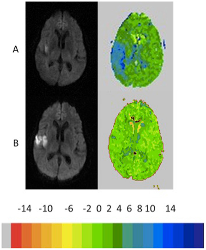
Diffusion-perfusion mismatch in sub-six hour acute stroke. (A) DWI... |  Download Scientific Diagram
Diffusion-perfusion mismatch in sub-six hour acute stroke. (A) DWI... | Download Scientific Diagram

Frontiers | Diffusion–Perfusion Mismatch: An Opportunity for Improvement in  Cortical Function
Frontiers | Diffusion–Perfusion Mismatch: An Opportunity for Improvement in Cortical Function

Amide proton transfer imaging in stroke - Heo - 2023 - NMR in Biomedicine -  Wiley Online Library
Amide proton transfer imaging in stroke - Heo - 2023 - NMR in Biomedicine - Wiley Online Library

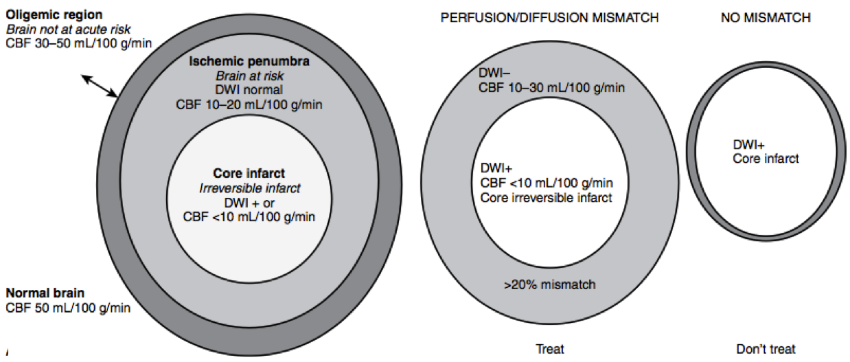
Magnetic resonance diffusion-perfusion mismatch in acute ischemic stroke:  An update
Magnetic resonance diffusion-perfusion mismatch in acute ischemic stroke: An update

Figure 2 from Salvage of the PWI/DWI Mismatch up to 48 h from Stroke Onset  Leads to Favorable Clinical Outcome | Semantic Scholar
Figure 2 from Salvage of the PWI/DWI Mismatch up to 48 h from Stroke Onset Leads to Favorable Clinical Outcome | Semantic Scholar
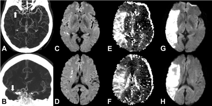
Rapid identification of a major diffusion/perfusion mismatch in distal  internal carotid artery or middle cerebral artery ischemic stroke | BMC  Neurology | Full Text
Rapid identification of a major diffusion/perfusion mismatch in distal internal carotid artery or middle cerebral artery ischemic stroke | BMC Neurology | Full Text

Rapid Assessment of Perfusion–Diffusion Mismatch | Stroke

EPOS™
EPOS™

Patient No. 10; CT perfusion showed mismatch in the right middle... |  Download Scientific Diagram
Patient No. 10; CT perfusion showed mismatch in the right middle... | Download Scientific Diagram

Estimating Perfusion Deficits in Acute Stroke Patients Without Perfusion  Imaging | Stroke
Estimating Perfusion Deficits in Acute Stroke Patients Without Perfusion Imaging | Stroke

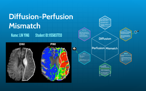
Diffusion-Perfusion mismarch by ying lin on Prezi Next

Salvage of the PWI/DWI Mismatch up to 48 h from Stroke Onset Leads to  Favorable Clinical Outcome - H. Ma, P. Wright, L. Allport, T. G. Phan, L.  Churilov, J. Ly, J. A.
Salvage of the PWI/DWI Mismatch up to 48 h from Stroke Onset Leads to Favorable Clinical Outcome - H. Ma, P. Wright, L. Allport, T. G. Phan, L. Churilov, J. Ly, J. A.

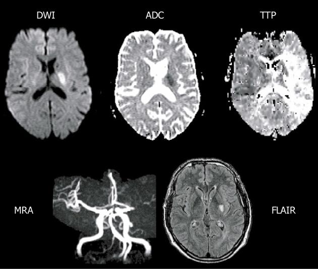
Acute arterial ischemic stroke with diffusion–perfusion mismatch.a |... |  Download Scientific Diagram
Acute arterial ischemic stroke with diffusion–perfusion mismatch.a |... | Download Scientific Diagram
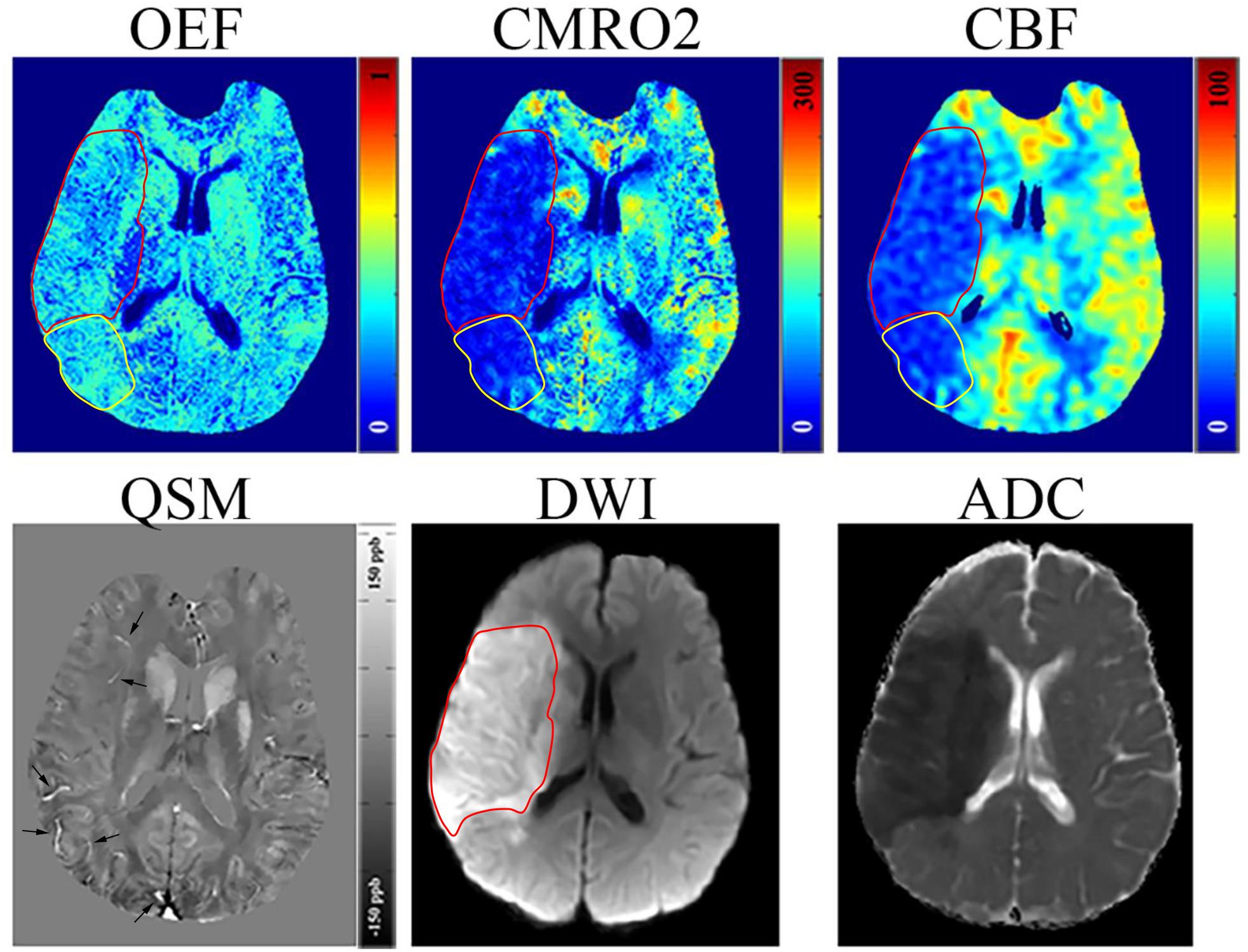
Frontiers | Initial Experience of Challenge-Free MRI-Based Oxygen  Extraction Fraction Mapping of Ischemic Stroke at Various Stages:  Comparison With Perfusion and Diffusion Mapping
Frontiers | Initial Experience of Challenge-Free MRI-Based Oxygen Extraction Fraction Mapping of Ischemic Stroke at Various Stages: Comparison With Perfusion and Diffusion Mapping

The diffusion-perfusion mismatch. A small lesion on the diffusion... |  Download Scientific Diagram
The diffusion-perfusion mismatch. A small lesion on the diffusion... | Download Scientific Diagram

MRI software for diffusion-perfusion mismatch analysis may impact on  patients' selection and clinical outcome | European Radiology
MRI software for diffusion-perfusion mismatch analysis may impact on patients' selection and clinical outcome | European Radiology
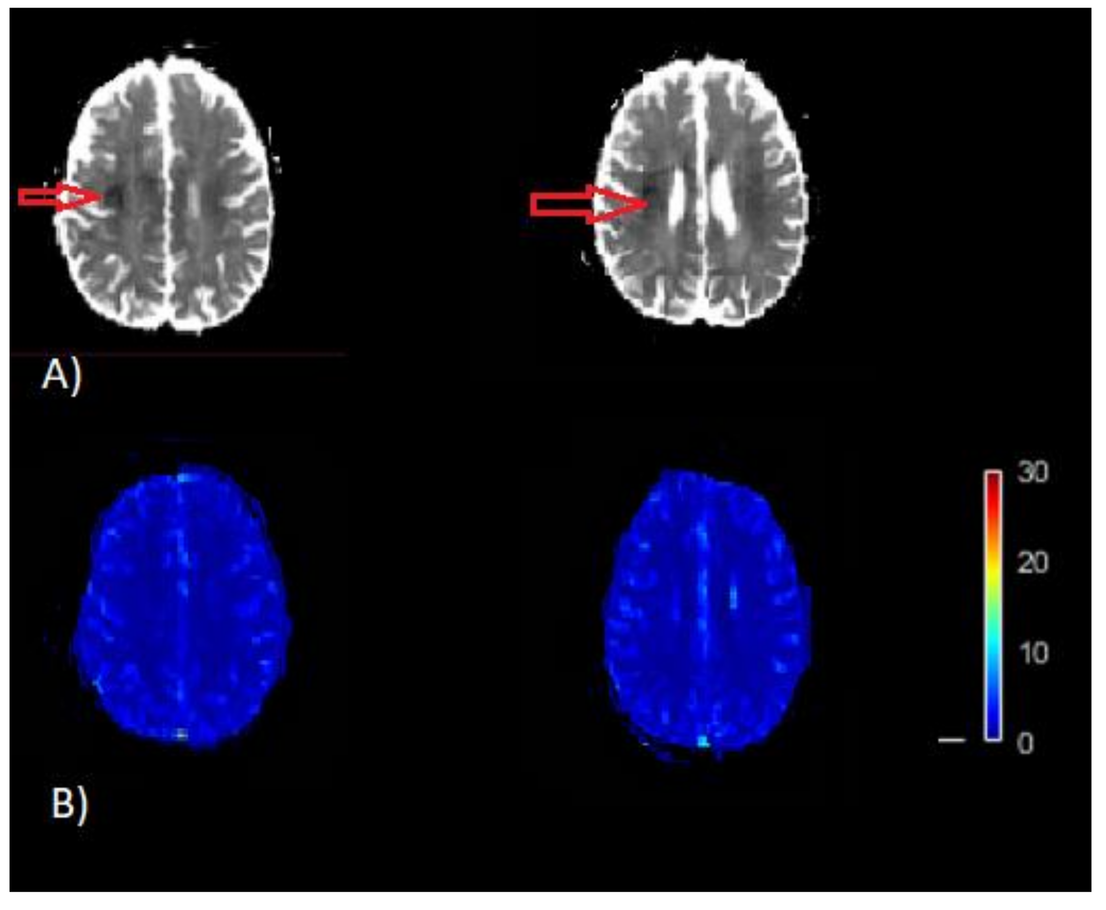
Brain Sciences | Free Full-Text | Optimal Scaling Approaches for Perfusion  MRI with Distorted Arterial Input Function (AIF) in Patients with Ischemic  Stroke
Brain Sciences | Free Full-Text | Optimal Scaling Approaches for Perfusion MRI with Distorted Arterial Input Function (AIF) in Patients with Ischemic Stroke
The Radiology Assistant : Imaging in Acute Stroke

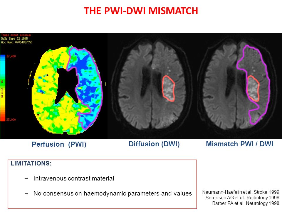
Ischemic penumbra in acute MCA stroke: comparison of the PWI-DWI mismatch  and the ADC-based Neurinfarct methods Drier A 1, Tourdias T 2, Attal Y 3, -  ppt video online download
Ischemic penumbra in acute MCA stroke: comparison of the PWI-DWI mismatch and the ADC-based Neurinfarct methods Drier A 1, Tourdias T 2, Attal Y 3, - ppt video online download

Diffusion- Susceptibility Mismatch in Stroke - YouTube

Figure 1 from Existence of the diffusion-perfusion mismatch within 24 hours  after onset of acute stroke: dependence on proximal arterial occlusion. |  Semantic Scholar
Figure 1 from Existence of the diffusion-perfusion mismatch within 24 hours after onset of acute stroke: dependence on proximal arterial occlusion. | Semantic Scholar

Diffusion–perfusion mismatch in sub-six hour acute stroke. - ppt download

Diffusion-perfusion mismatch indicating viable ischemic brain tissue... |  Download Scientific Diagram
Diffusion-perfusion mismatch indicating viable ischemic brain tissue... | Download Scientific Diagram

Can diffusion- and perfusion-weighted imaging alone accurately triage  anterior circulation acute ischemic stroke patients to endovascular  therapy? | Journal of NeuroInterventional Surgery
Can diffusion- and perfusion-weighted imaging alone accurately triage anterior circulation acute ischemic stroke patients to endovascular therapy? | Journal of NeuroInterventional Surgery

Diffusion- and Perfusion-Weighted Imaging in Acute Lacunar Infarction: Is  There a Mismatch? | PLOS ONE
Diffusion- and Perfusion-Weighted Imaging in Acute Lacunar Infarction: Is There a Mismatch? | PLOS ONE

Beyond Mismatch | Stroke

Automated MRI perfusion-diffusion mismatch estimation may be significantly  different in individual patients when using different
Automated MRI perfusion-diffusion mismatch estimation may be significantly different in individual patients when using different